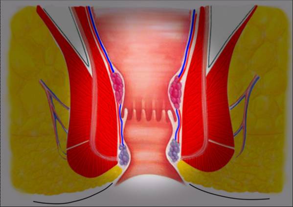
It is one or more absences, acute or chronic, with drainage of pus and blood in the sacrococcox area.
INDEX:
1. WHAT IS THE SACROCOCCYGEAN FISTULA
2. TREATMENT OF SACROCOCCIGEAN FISTULA
3. SURGICAL TECHNIQUES
3.1. MINIINVASIVE TECHNIQUE EPSIT
3.2. OPEN TECHNOLOGY
3.3. CLOSED TECHNIQUE
4. GENERAL INFORMATION AFTER THE INTERVENTION FOR PILONIDALIS CISTS OR FISTULA
1. WHAT IS THE SACROCOCCIGEAL FISTULA
It is also called CYST or PILONIDAL FISTULA and also SINUS PILONIDALIS.
It is one or more abcesses, acute or chronic, with drainage of pus and blood in the sacrococcigeal area.
It also manifests itself with pain, swelling, skin redness, fever. It is most common in males (3: 1 M: F) predominantly between 15 and 40 years.
While in the past it was believed to be a congenital disease, it is currently believed that there is an infection of the hair follicles in that area.
The formation of an abscess would be followed by a push of the surrounding skin into the abscess cavity due to the clutch generated during the movement of the buttocks muscles in the walk.
If untreated, it can evolve into multiple fistulas, ie openings to the outside, with various abscesses. Seldom described, though very rarely, malignant degeneration of untreated relapsing sacrococcus abscesses.
2. TREATMENT OF SACROCOCCASE FISTULA
Sacrococcuse abscesses can be drained into local anesthesia.
The release of pus and hair inside cysts can be the ultimate cure in 60% of the cases.
If the drainage of the material from cysts continues within three months, a definitive treatment is required.
Weekly depilation of the area, followed by accurate local hygiene reduces the risk of recurrence of 90%.
3. SURGICAL TECHNIQUES
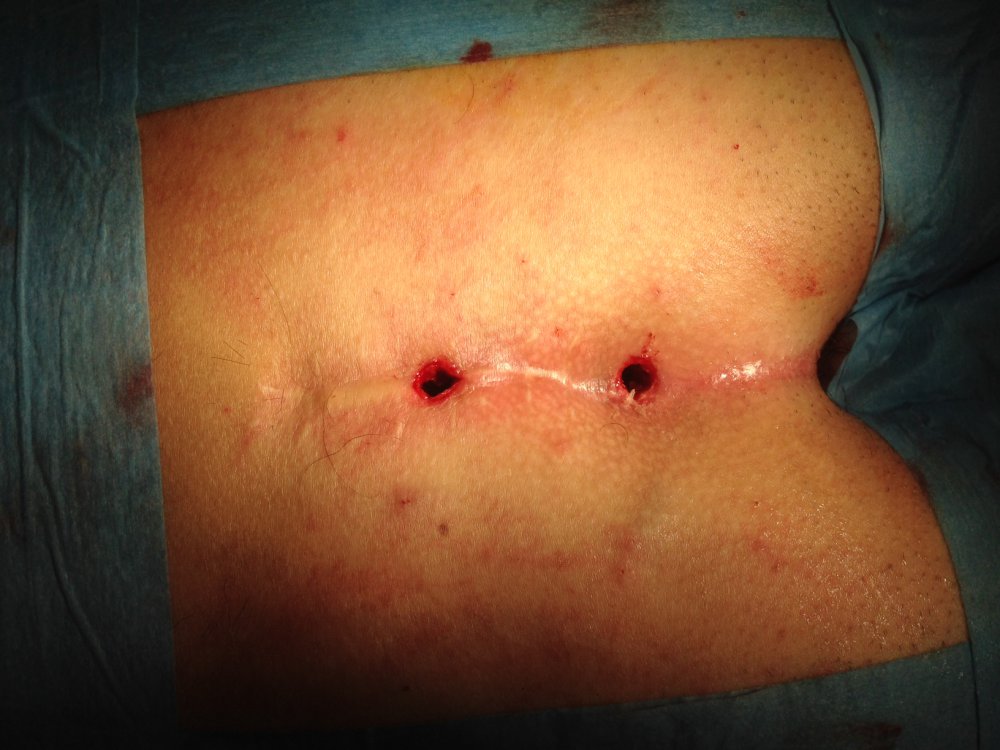
In most cases, surgery is performed under Day Hospital, ie without spending the night at the Hospital and can be done in several ways by surgically removing the inflamed area. In recent years there is growing interest in a MINIINVASIVE method called EPSIT (Endoscopic Pilonidalis SInus Treatment) that I currently recommend in many cases.
3.1 MINIINVASIVE TECHNIQUES - EPSIT (Endoscopic Pilonidal Sinus Treatment)
This is a revolutionary, mini-invasive Video Assistance technique, conceived by an excellent Italian surgeon, Dr. Meinero.
The search for a less obnoxious method results from the fact that all the other techniques create a long, painful postoperative course with unexpected scars that, especially in women, create little embarrassment.
In simple words, it is about creating or widening the natural hole of the fistula up to about 5 mm and penetrating the "tunnel" of the fistula or cavity cavity with a particular instrument (FISTULOSCOPE) containing a miniature camera, that is, very small, A washing channel and an operator channel where an electrocoagulator, or a clamp can be introduced. the
Images are displayed and enlarged on a screen.
Penetrating inside the fistula, the infected area, the hair masses, typical of this disease, and the various branches of the sinus appear.
Then remove the contents of the cyst and the fistula, and with the appropriate electrocoagulator it cauterizes, that is, the walls are burnt.
The aesthetic result is exceptional. Relapses overlapping with those with other methods. Seldom there is postoperative pain treated with painkillers for a couple of days.
The resumption of work or schooling will be a few days.
3.2 OPEN TECNIQUE
Escaping the cysts and praising the cavity with gauze followed by biweekly medications until the complete defect closure in one-two months. I do it more and more rarely and only in cases of relapsing illnesses with severe local infections
3.3 CLOSED TECHNIQUE
Escaping the cyst and closing it with a tissue plastic that reduce the tension.
Often a drainage tube is needed for a few days and can be performed in local anesthesia (anesthetic injections on the area). The intervention begins by inoculation of a dye in the outer aperture of the fistula, then it removes all the pathological tissue remaining in the area not soiled by the dye.
After coagulating the small blood vessels in the wound, the wound is closed with multiple layers of dots.
If the removed tissue is too large, a cute and subcutaneous area should be prepared and a so-called rotating flap must be made to close the defect without risk of excessive tissue tension.
The post-operative course involves a forced rest and a difficulty sitting for about fifteen days.
4. General information for the patient after surgery for SACROCCIGEA fistula with miniinvasive technique E.P.S.I.T.
After the intervention, the area remains painless thanks to a fixed-dose and time-stamping therapy that will continue to be home and allow for a new life

.JPG?box=75x75c)


.JPG)