The breast cancer is the most common cancer in the woman, although at present and thanks to the goals it has achieved in therapeutic research, it no longer represents the main cause of cancer death. It affects women at any age with a peak around the age of 60.
• MOST AFFECTED :
There is a higher incidence in women who have not had pregnancies, in those with late pregnancies (over 35), in those with a family history of breast cancer or with a personal history of breast cancer.
• HOW MANIFESTED:
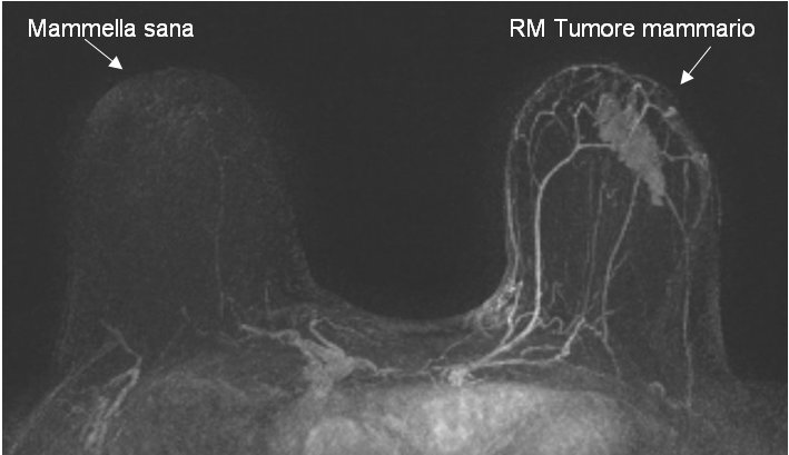
If palpable you appreciate a hard mass, fixed and with margins not defined; If not palpable, mammography and ultrasound will recognize it even at a very early stage.
• CONSEQUENCES:
Ulcerations, abnormal increase in axillary lymph nodes and over the clavicle, arm edema, remote metastases such as bone, lung, liver and brain.
• SURGICAL TECHNIQUES:
Most women who come to my observation are carriers of early tumors and already have a cytological or histological diagnosis that confirms their nature.
Approximately 10 years ago, important studies have been published (both in Milan and in the United States) that demonstrated that with the simple removal of tumor mass and axillary lymph nodes followed by radiotherapy, the same results were obtained for the complete removal of the breast and axillary lymph nodes in Free period of illness and long-term survival.
Therefore, the tendency where possible is to perform surgery aimed at removing the tumor with an adequate margin of healthy tissue and not that of removing the entire breast which is reserved for cases of locally advanced or multicenter tumors.
The axillary lymph nodes are those that receive the lymph from the breast and therefore also from the tumor.
When tumor cells break away from breast cancer, they travel to the lymph and stop in the axillary lymph nodes where they reproduce by increasing the lymph nodes (lymph node metastases).
Knowledge of the number of lymph nodes involved in the tumor is an important indicator to determine the severity of the disease and to establish the correct therapy to be followed after the intervention.
This is why it always associates the removal of axillary lymph nodes to breast surgery.
As only 30 breast cancer out of 100 have axillary lymph node metastases, 70 women per 100 receive axillary lymphadenectomy without any need. Always with the aim of ensuring the best result in healing with the slightest possible trauma, other studies have shown that the mere removal of lymph node that first receives lymph from the cancer, the so-called "sentinel lymph node" is a good indicator of involvement or Less than the other axillary lymph nodes.
Therefore, the scintigraphic technique of the removal of the sentinel lymph node has become a foundation of breast surgery and to be implemented besides the experience of the surgeon also of an adequately organized structure with nuclear medicine.
The woman who is involved in tumor or quadrant surgery (removal of a breast tissue part corresponding to a dial) with sentinel lymph node, the day of the intervention or the first one, will be present at the nuclear medicine center under the ultrasound guidance, He will be injected with a tumorous needle, a marker like technetium.
After two hours, the armpit is checked with a camera range and the area where the technetium has accumulated is identified by marking it on the skin with an indelible marker.
Often, when the tumor is not palpable, I use the same scintigraphic technique to identify it at the operative site.
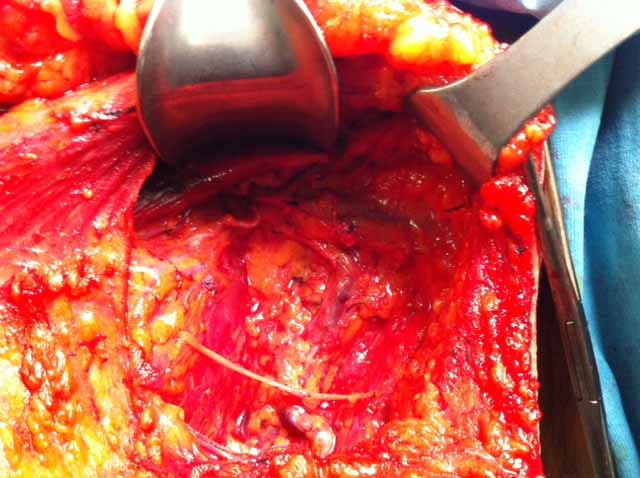
The surgery is performed in general anesthesia or in local anesthesia with sedation.
With the help of a probe that detects the gamma rays of the marker injected earlier, I can do a targeted removal of the tumor with a suitable free surrounding margin.
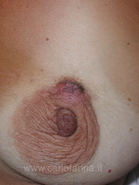
When it is possible the engraving will respect the aesthetics of the breast and will tend to be inherited from the dark area of the lobster and the clear breast.
Depending on the amount of tissue removed, I place a small drainage tube and close the engraving with dots not visible on the skin or emerging only in the area of the crown so as not to leave the mark (Algower points).
Practical then another small engraving in the area of the aisle marked with the pen marker and under the guidance of the detector probe I take only the lymph nodes I am signaling.
The removed tumor and sentinel lymph node are sent to the anatomopathologist for histological evaluation. If the lymph node does not contain tumor cells the intervention is terminated otherwise it will re-intervene to remove all the axillary glands after about 20-30 days.